Urethral Strictures

kidney stone disease 1

What is stricture?

  • In males the urethra starts from the outlet of the bladder and runs through prostate gland .This part of the urethra is called the posterior part. Then it becomes broader (Bulbar Urethra) and then It continues through the length of penis (Anterior urethra) and exits through a opening called urethral meatus.
  • The urethra has a lining and is surrounded by specialized spongy tissue which supports the urethra when the lumen of the urethra is narrowed the condition is called urethral stricture. The anterior urethra is very much prone to inflammation or direct injury.
  • The posterior urethra is more or less fixed and can be damaged during major injury resulting in fracture of pelvic bones.
  • Urethral stricture disease has been cited as long ago as ancient Greek writings that report establishing bladder drainage with the passage of various catheters. Historically, the treatment consisted of urethral dilation with sounds. In modern days better treatment options are available with better outcome.

What causes the urethral stricture?

  • Any process that injures the urethral lining and the surrounding corpus spongy tissue ultimately heals in scar can cause urethral stricture.
  • The most common causes of urethral stricture today is following injury during road traffic accident.
  • Sometimes direct injury to the area happens and the patient hardly can remember the episode. This can lead to stricture at a later date.
  • Urethra can also get affected during passage of endoscopy for investigation and treatment.
  • Venereal infection such as Gonocoocal infection causing stricture is fortunately rare today.
  • Sometimes scarring of tip of the penis due to inflammation can involve urethral opening and stricture can extend into the urethra (Lichen sclerosis).

What symptoms does the patient have?

  • Obstructive voiding symptoms are characterized by decreased force of stream, incomplete emptying of the bladder, terminal dribbling, and interrupted urinary stream.
  • Symptoms of urinary tract infections.(Fever, painful and urination).
  • Infection affecting the epididymis presenting as scrotal swelling.
  • Sudden stoppage of urine (urinary retention).

What happens when treatment is delayed?

  • Poor quality of life with frequent urination, cannot go to places.
  • Chronic urinary retention with overflow incontinence.
  • Urine leaking into the skin (Urethrocutaneous fistula).
  • Reduction in kidney function (renal failure).

How is the diagnosis made?

The following tests are done to confirm the diagnosis as well as the length of stricture to plan the treatment

  • Urine flow test.
  • The speed at which urine passed can be assessed by simple uroflowmeter. The patient has to pass urine when the bladder is full .urine flow rate below 20 ml/ sec is suggestive of obstruction in the urethral passage.
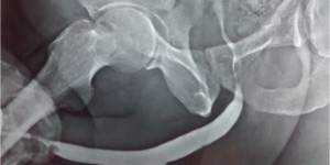
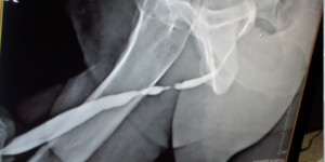
  • X-ray studies, Ascending urethrogram.
  • This test involves placing a fine catheter into the urethral opening and injecting contrast media .The narrowed segment and the length of narrowed segment can be visualized.
  • Endoscopic evaluation can be conducted by flexible or rigid instruments (cystourethroscopy). Flexible cystourethroscopy can be performed with little discomfort to the patient using only local anesthesia.

How can we treat this problem ?

1. Urethral Dilatation :

The stricture can be dilated using special dilators to stretch the narrowed area and make it wider. This usually requires to be repeated at regular intervals to keep the passage wider. Complications include recurrence of stricture, which is the most common complication, Success with its treatment is very poor 15% only.

2. Internal urethrotomy (Optical urethrotomy) :

  • Internal urethrotomy involves incising the stricture transurethrally using endoscopic equipment. The incision allows release of scar tissue. Success depends on the epithelialization process finishing before wound contraction significantly reduces the urethral lumen caliber. The incision is made under direct vision at the 12 o'clock position with an instrument called urethrotome. There is no open incision cutting and the procedure is entirely endoscopic. . Typically, an indwelling urethral catheter is left in place for 3-5 days to oppose wound contraction forces and allow epithelialization.
  • The curative success rate is reported as 20-35%, with no increase in the success rate with repeating the procedure.

3. Open reconstruction (Urethroplasty) :

  • A Primary repair involves complete excision of the fibrotic urethral segment and joining normal looking segments. (Excision Urethroplasty). There should not be any tension at the repair site. This method is suitable only for stricture lengths of 1-2 cm. With extensive mobilization of the corpus spongiosum, strictures 3-4 cm in length can be repaired using this technique. This gives excellent success rate.
  • If the narrow area is longer than 3 cm and if the stricture location within the penile shaft , the gap cannot be bridged after excising the affected segment .This can be managed by one of the two methods.
  • In one method , after excision of narrowed segment and interposing vascularised tissue from penile skin, scorotal skin or lining from the cheek (Buccal mucosa) and reconstructing urethra with normal diameter.
  • If the stricture length is long, the the entire narrow segment is opened and long graft from inside the cheek is placed and joined to the opened urethra on its side to make the urethra diameter wider. After good healing of this site, second procedure is done to close the urethral lumen and the surgery is completed (two stage urethroplasty).