Kidney Stone Disease

kidney stone disease 1

About Kidney Stone

  • Urine is filtered from blood by glomerulus, basic filtering unit of kidney. The calyces, renal pelvis form the drainage tubes. The calyces are divided into upper, middle and lower parts. Stone are formed at the calyceal level and it may increase in size, and or move down to ureter via renal pelvis. If it can reach the bladder it is usually passed out in the urine. Sometimes it can get stuck within the renal calyces, renal pelvis, ureter or urethra.
  • Person taking high intake of animal protein and reduced intake of water, those with obstruction to urinary system drainage and having residual urine are also prone to stone formation.
  • There is a genetic element also and family members can have stone.

Types Of Stones :

  • Calcium and oxalate stones are the commonest.
  • Uric acid stones are less easily visible on x-ray.
  • Triple phosphate stones are remain symptomatic and invariably stones are bigger in size.
  • Staghorn stone ( large stone, with branching occupying part or most of the pelvicalyceal system.

Size Of Stone :

  • 3 to 5 mm stones are considered small. These stones usually get passed in the urine in over 80% of the time with plenty of fluid and medication. Rarely even these small stones need endoscopy to remove them if they are stuck and do not move.
  • 7 to 10 mm stones are considered medium size stone. Spontaneous passage of stone is around 70%.
  • Stones of 1 cm or more considered as large in stone and only 30% chance of spontaneous passage can be expected.

How do I Know Whether Stone is Passed ?

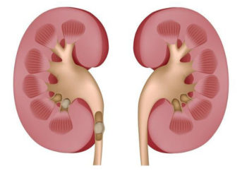
One should see stone in the urine that is passed. Absence of pain does not mean that stone is passed, stone can remain and cause damage to the kidney. Presence of stone has to be checked by x-ray, scan or both.

Stone In Single Kidney : Needs Urgent Treatment

  • Some people are born with one kidney only instead of normal two (1 in 5000).
  • If one had already have one kidney with poor function or one kidney removed due to disease, then presence of stone in a single functioning kidney needs great attention.
  • Patient must pass the stone early or the stone should be removed at the earliest to restore function.

Symptoms Of Stone Disease :

  • Pain in the side of abdomen (loin pain).
  • Blood in the urine.
  • Fever and vomiting.
  • Difficulty in passing urine.
  • Infected stones may remain asymptomatic and can cause mild back pain.

How Is The Diagnosis Confirmed?

  • Blood tests are done to confirm the kidney function and indicators for urinary infection.
  • Ultrasound scan, CT scan and x-rays are usually adequate to confirm the presence of stone, it's location and size and severity of obstruction.

What Complications occur If not Treated In Time ?

  • Infection in the kidney (Pyonephrosis- pus collection).
  • Leading to loss of kidney function (Kidney failure).

How Needs Endoscopic Surgery For Stones?

  • If stone is not passed after reasonable period of time and causes pain.
  • Stone is too large to pass on it's own.
  • Stone associated with severe obstruction.
  • Reduction in kidney function is a sign of urgent need to stone removal.

Types Of Endoscopic Procedures :

  • Cystoscopy and laser for bladder stones.
  • Rigid ureteroscopy and laser treatment.
  • Flexible ureteroscopy and laser treatment.
  • Percutaneous nephrolithotomy using laser.

Cystoscopy and Laser :

  • An instrument called Cystoscope passed small and medium are removed by special instrument called Lithotrite.
  • Large stone are powdered using laser energy.

Large Bladder Stone

Rigid Ureteroscopy and Laser :

  • Stones within the ureter are treated using Rigid ureteroscopy.
  • Long thin scope can be passed upto the level of stone in the ureter. Small stone is completely removed with forceps and larger ones require fragmentation using pneumatic lithotripsy. Dr. Anandan uses Laser for fragmentation of stone using dusting technique where stones are powdered and do not migrate up during the procedure. Urine that passes pushes the fragments in to the bladder and are then passed out in the urine.
  • A stent may be required to provide good drainage of urine.

Retrograde Intrarenal Surgery (RIRS):

  • Retrograde Intrarenal Surgery (RIRS) and Laser Lithotripsy for stones within the kidney.
  • Specially designed scope to look and treat stones within the kidney is called Flexible Ureteroscopy.
  • The tip of the flexible ureteroscope can bend both up and down in a single plane and allows easy access to the calyceal system which is not possible with rigid scope.There is a channel in the center of scope through laser fibre for breaking the stone, baskets and similar retrieval devices can be passed. Current flexible ureteroscope are small in diameter, better optics and capable of visualising the calyces in 94 to 100 % of cases. Laser can break stones of very hard nature as well.
  • Ureteroscopy is one of the common procedures used to treat kidney stones. It is also used to diagnose and treat a variety of conditions such as polyps or cancer in the urinary tract. Cancers can also be treated in the early stages with laser.
  • It takes about one hour to complete the procedure, which may or may not require an overnight stay. A self retaining tube , one end of which will fold itself in the kidney and the other end will coil itself in the bladder called ureteric stent will be placed. This allows stone fragments to pass down the ureter in to the bladder and then patients passes stone fragments in the urine It also relieves obstruction to the urine flow. A repeat Scan or x-ray will assess completeness of stone clearance. The stent is removed 2 to 4 weeks later as a day case procedure.

Flexible Ureteroscope

Stone in the Kidney treated by Laser

Laser Machine

PCNL ( Mini Percutaneous Nephrolithotomy) :

  • If the stone volume is large ,it cannot be effectively managed by lithotripter.
  • A tract is created from the back to the collecting system containing the stone, Nephroscope is passed and the stone is visualized. Broken with ultrasound or laser probe. As the tract is small there is minimal discomfort at the site of surgery and it heals well.
  • Generally one, but rarely more punctures tract are needed to clear the stone.
  • There is generally one single 1 cm cut for this procedure.